AFK weekly newsletter 3

Jul 20 / Mohamed Moussa
Did you diligently prepare for the upcoming assessment of fundamental knowledge exam (AFK)? Are you seeking a valuable resource that provides top-notch questions accompanied by detailed explanations? Look no further than this exclusive weekly newsletter tailored just for you. Our dedicated community commits to delivering a thought-provoking question each day to stimulate your intellectual prowess. But that's not all! As the weekend approaches, we go the extra mile by creating engaging videos that not only provide the correct answers but also delve into the underlying concepts behind them. Immerse yourself in the rich learning experience we have curated for you. Whether you prefer watching the informative videos or simply perusing the carefully crafted questions and answers, we have got you covered. Join our community now and embark on a journey towards mastering the fundamental knowledge required for your success!

AFK Weekly newsletter 3

In just 30 minutes, you will get some really important concepts for AFK. 
  • Explain the answers in details. 
  • Quick recap for the whole concept.
Write your awesome label here.

#Operative
1-increasing the volume of fillers in composite resin will cause all of the following, except:
◯ a. increased hardness or strength
◯ b. Increase polymerization shrinkage
◯ c. lower thermal expansion coefficient
◯ d. increase of compression strength
 b. Increase polymerization shrinkage
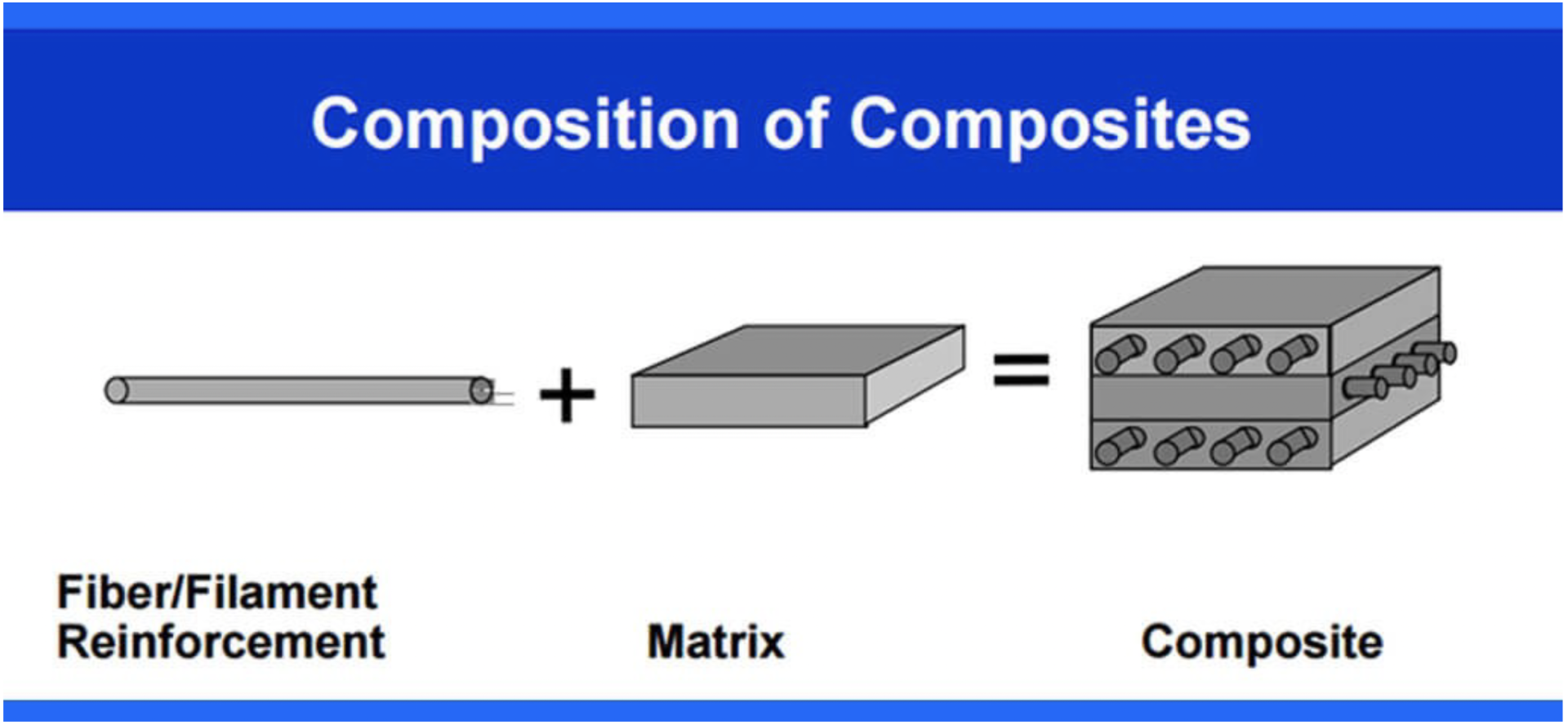
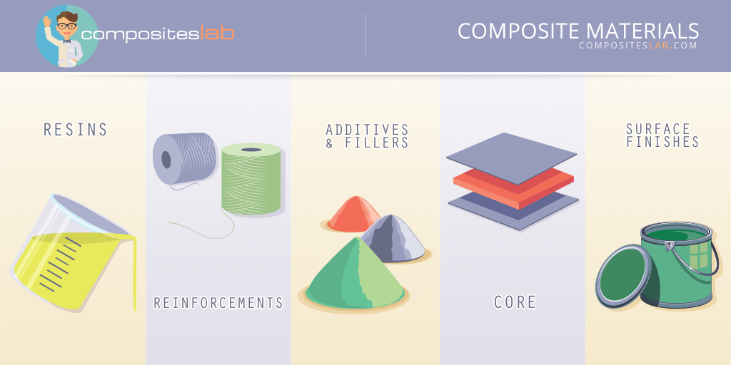
- Dental composites are composed of several main components, let's discuss the different components. Then explain in details about the function of the fillers in dental composite. 

1- Organic Matrix:

The organic matrix forms the bulk of the dental composite and is responsible for its physical and mechanical properties. It consists of a resin matrix, typically composed of bisphenol A-glycidyl methacrylate (Bis-GMA), urethane dimethacrylate (UDMA), or other methacrylate-based monomers. The resin matrix provides the composite with its polymerizable and adhesive properties.

2- Fillers:

Fillers are added to the organic matrix to enhance the composite's strength, wear resistance, and aesthetic properties. They can be inorganic particles such as glass, quartz, or ceramic particles. The fillers reinforce the composite and give it the ability to withstand chewing forces. They also provide the desired color and translucency to mimic natural teeth.

Other components

 are Coupling Agents, Initiators and Accelerators and Pigments.

- Function of the fillers in the composite are:
1- Reinforcement:

Fillers are added to composite materials to enhance their mechanical properties and improve their strength. The fillers reinforce the composite matrix, making it more resistant to wear and fracture.

2- Wear resistance:

Fillers help increase the wear resistance of dental composites, making them more durable when subjected to chewing forces and abrasive actions.

3- Aesthetic properties:

Fillers are responsible for mimicking the natural appearance of teeth. They provide opacity and color matching capabilities to dental composites, allowing them to blend seamlessly with the surrounding tooth structure.

4- Polymerization control:

Fillers play a role in controlling the polymerization process of dental composites. They help to regulate the shrinkage and curing characteristics, reducing the polymerization stress that can lead to marginal gaps or tooth sensitivity.

5- Handling properties:

Handling properties: Fillers can influence the viscosity and flowability of dental composites, making them easier to handle during placement. The fillers provide the necessary rheological properties for shaping and contouring the composite material.

6- Radiopacity:

Some dental fillers contain radiopaque materials, such as barium, strontium, or zirconium, which make the composite visible on X-rays. This radiopacity enables dentists to monitor the restoration's integrity and diagnose any potential issues.

#Pharma
2-A patient is taking Atenolol, Ososorbide Mononitrate and Diltiazem (Cardiazem). The most likely diagnosis is:
◯ A. Atrial fibrillation and congestive heart failure
◯ B. Congestive heart failure and myocardial infarction
◯ C. Cardiac ischemic disease and hypertension
◯ D. Myocardial infarction and ventricular arrhythmia

 C. Cardiac ischemic disease and hypertension
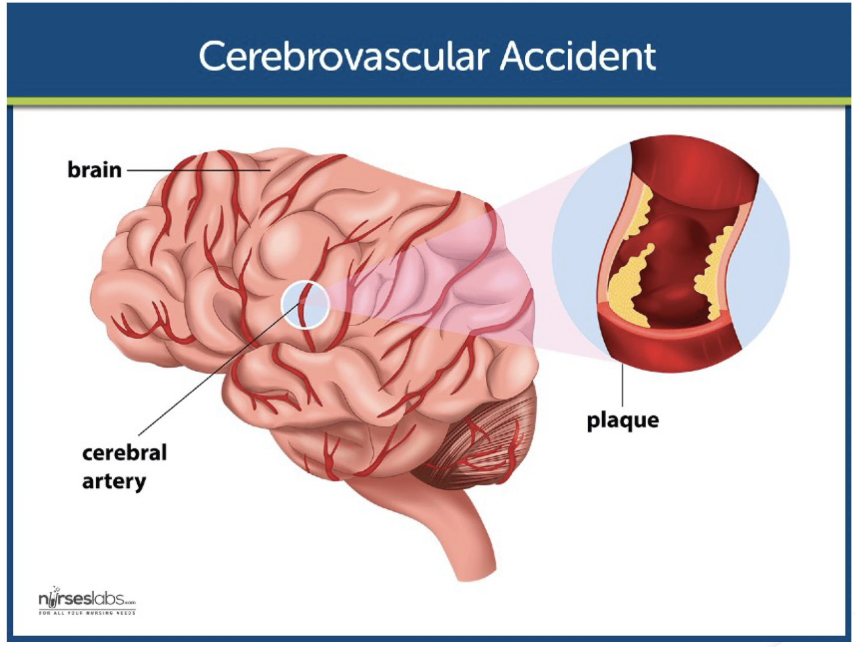
- Cerebrovascular Accident
Anticoagulants
(heparin or warfarin)
Antiplatelet agents (aspirin or clopidogrel).
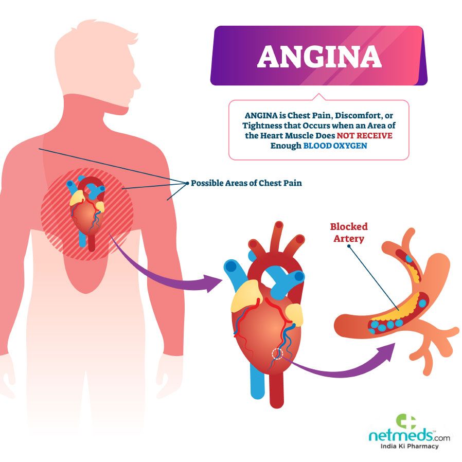
Stable Angina:
Nitrates,
anti-platelets,
statins,
beta blockers,
CCBs,
ACE inhibitors,
 anti-platelet agents.
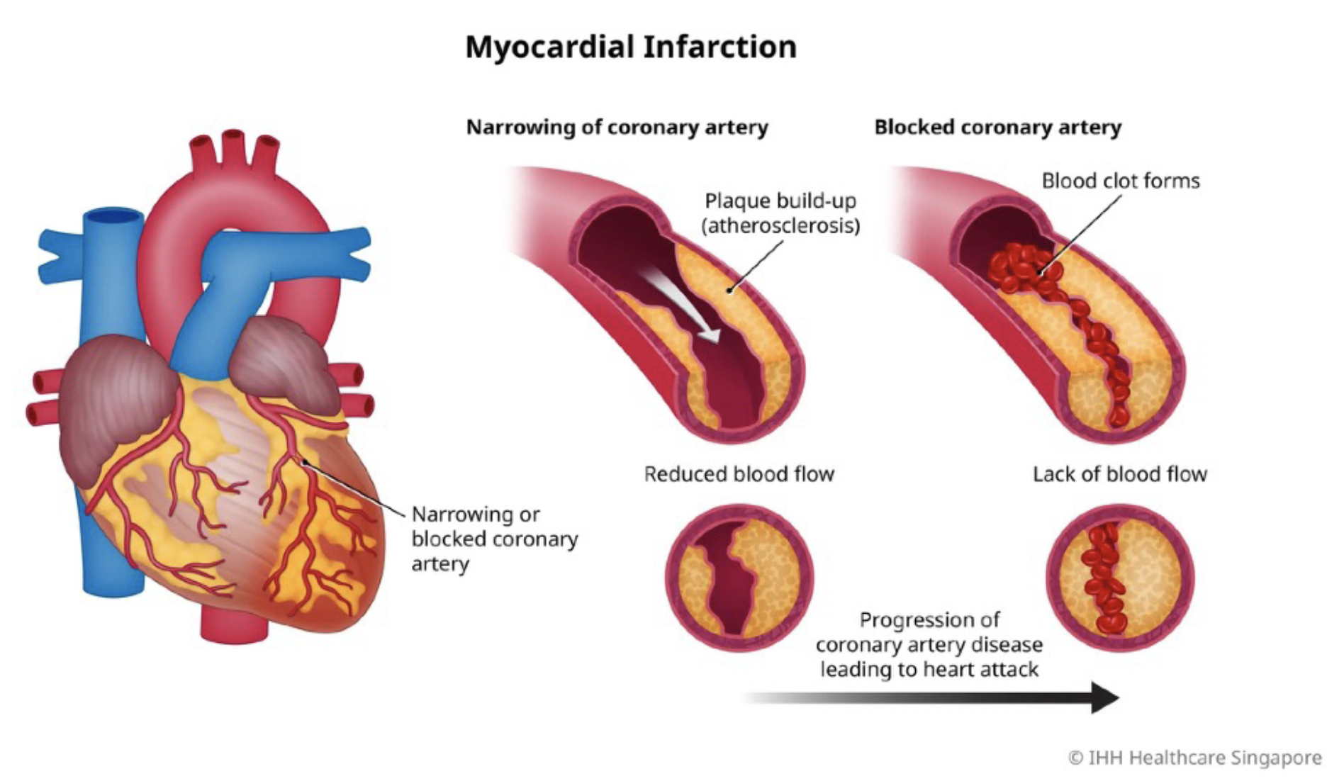
- Myocardial Infarction:
Same as angina
anticoagulants 
sedatives
morphine(Morphine and sedatives mainly used during acute MI).
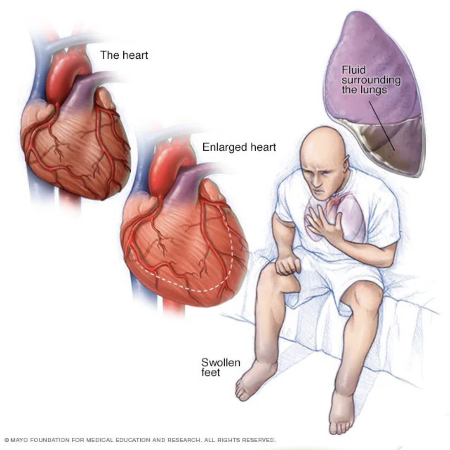
Congestive Heart Failure :
Diuretics,
Digitalis,
ARB's,
ACEl's,
beta blockers,
aldosterone antagonists,
nitrates.

Join our AFK Weekly newsletter

 Solving questions is really important, but solving exam grade questions with true and authenticated answers is critical.
Will post a question everyday, then at the week end will do a video to explain the correct answers and the concept of each questions, signup now to get weekly updates.
Thank you!

#Periodontology 
3- Chlorhexidine mouth rinses act on:
◯ A. Streptococcus mutans
 ◯ B. Lactobacilli
◯ C. streptococcus pyogenes
◯ D. Candida albicans

A. Streptococcus mutans
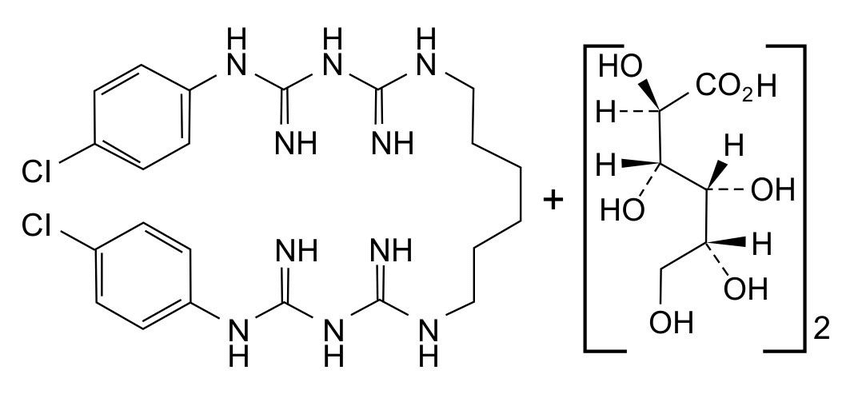
Chlorhexidine is a common antiseptic and antimicrobial agent
Chlorhexidine is a chemical compound that belongs to a group called bis biguanides. It's usually available as a liquid solution or gel, and it comes in different concentrations for different uses.
- Chlorhexidine Mode of Action: 
Chlorhexidine works by disrupting and damaging the outer membranes of microorganisms like bacteria, fungi, and viruses. It does this by binding to their cell walls, interfering with their integrity and preventing their growth and reproduction. This action makes Chlorhexidine an effective tool in fighting against oral infections.
- Chlorhexidine has several dental
- Prevention of Dental Plaque and Gingivitis.
- Chlorhexidine is commonly used as a mouthwash or oral rinse to control plaque buildup and prevent gingivitis (inflammation of the gums).
- It can inhibit the growth of bacteria in the mouth, reducing the risk of oral infections.

- Periodontal Maintenance: - In cases of periodontal disease, where there is infection and inflammation of the gums and supporting structures of the teeth, Chlorhexidine may be used as part of a comprehensive treatment plan to control bacterial growth and promote healing.

-Chlorhexidine (CHX) is effective against a wide range of bacteria, including both gram-positive and gram-negative species. Some of the bacteria that Chlorhexidine is particularly effective against include:

Streptococcus mutans:

This bacterium is a major contributor to dental plaque formation and tooth decay. Chlorhexidine can inhibit its growth, helping to prevent cavities.

Porphyromonas gingivalis:

This bacterium is associated with periodontal disease and gum inflammation. Chlorhexidine can effectively reduce its numbers, aiding in the treatment of periodontal conditions.

Aggregatibacter actinomycetemcomitans:

This bacterium is another common cause of periodontal disease. Chlorhexidine can help control its growth and prevent further damage to the gums and supporting structures of the teeth.

Prevotella intermedia:

This bacterium is associated with periodontal infections and can contribute to the progression of gum disease. Chlorhexidine has been shown to be effective against Prevotella intermedia.

Fusobacterium nucleatum:

This bacterium is commonly found in dental plaque and can contribute to gum disease. Chlorhexidine can inhibit its growth and reduce its harmful effects on oral health.

#Epidmology 
 4-Which of the following provides the best information according to evidence based dentistry?
◯ A. Case-Control studies
◯ B. Cohort studies
◯ C. Randomized clinical trails
◯ D. Systematic reviews
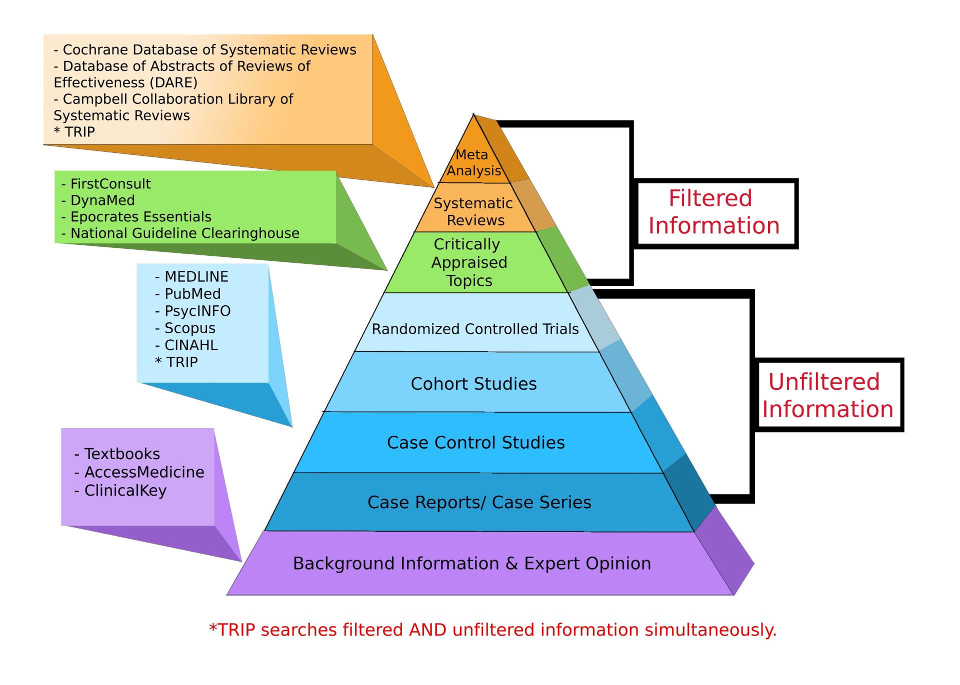
 D. Systematic reviews
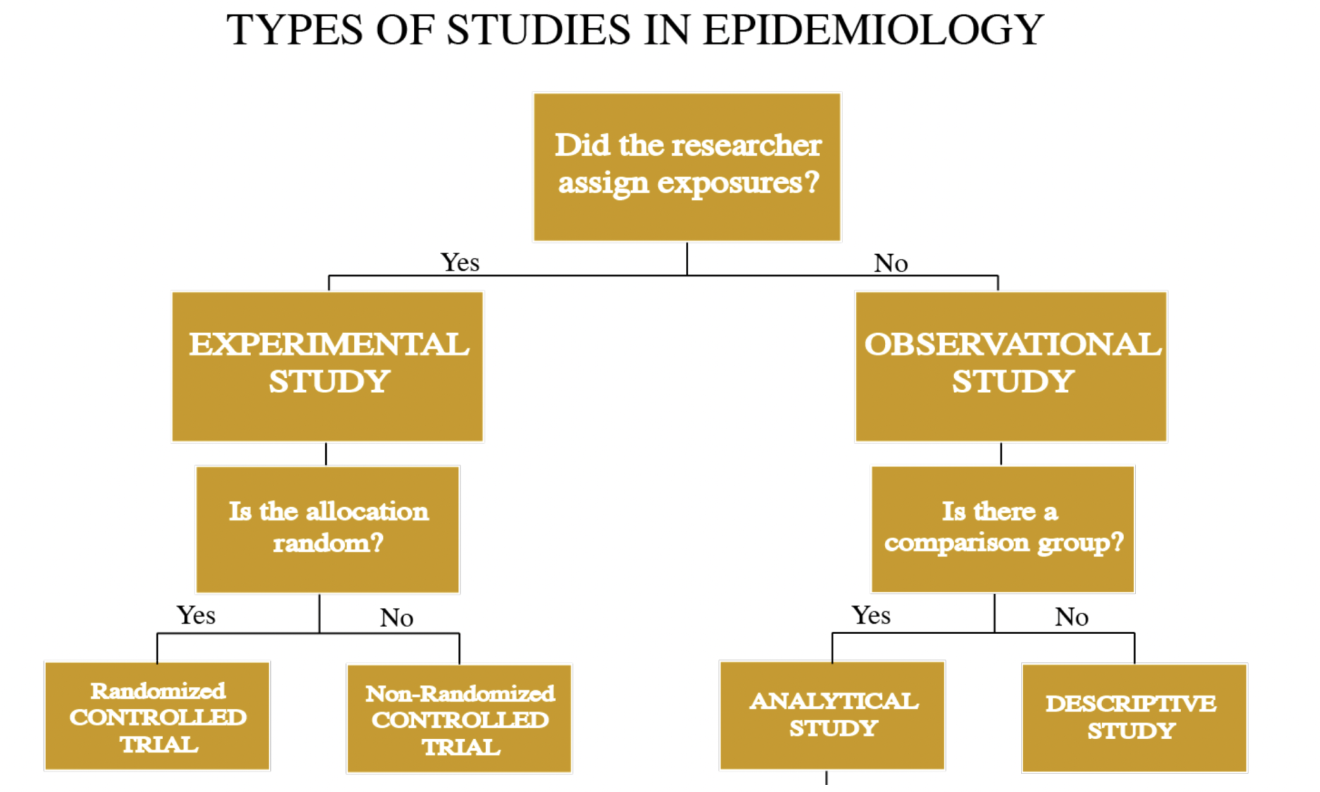
- Not all studies have the same credentials. Some studies like case report are just a documentation for some clinical cases.
 - There are 2 main types of studies
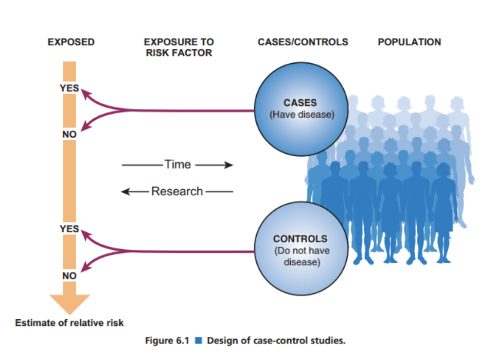
-Case-control will divide the subject to be one group with the disease (Cases). The other one will be without the disease (Control).
- Then will follow their exposure to different types of risk factors.
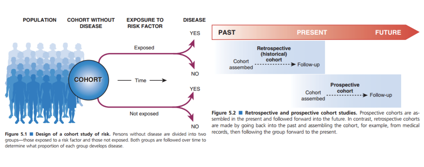
- Cohort will divided the subjects according to their exposure to risk factor, or not exposed.
- Then after years to check over if they developed the disease or not

#Periodontology 
5-A patient has a pocket depth of 6mm and attachment loss of 5.5 mm, what is the correct treatment in this case?
◯A. Scaling and root canal
◯ B. Oral hygiene measures
◯ C. Comprehensive treatment
◯ D. Surgical intervention

 D. Surgical intervention
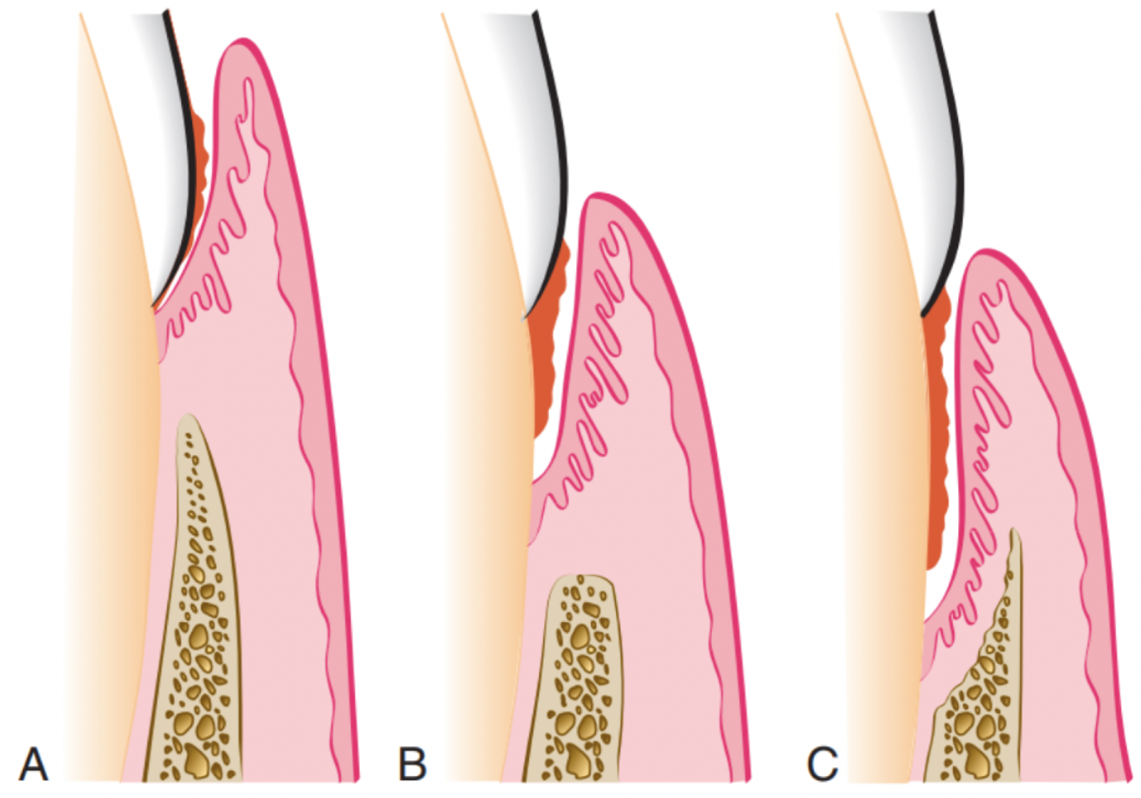
Different types of periodontal pockets. - (A) Gingival pocket.
There is no destruction of the supporting periodontal tissues.
- (B) Suprabony pocket.
 The base of the pocket is coronal to the level of the underlying bone. Bone loss is horizontal. -
(C) Intrabony pocket.
The base of the pocket is apical to the level of the adjacent bone. Bone loss is vertical.
Healing after any periodontal treatment will be either with repair like long junctional epithelium or with regeneration after periodontal surgery called guided tissue regeneration.

Phase I therapy / Rationale:

This Phase I therapy is a critical aspect of periodontal treatment.Data from clinical research indicate that the long-term success of periodontal surgical treatment is dependent on maintaining the plaque or biofilm control results achieved with phase I therapy.
- In fact, patients who do not have adequate plaque or biofilm control will continue to lose attachment regardless of what surgical procedures are performed.
- Based on the knowledge that microbial plaque or biofilm is the major etiologic agent in gingival inflammation, one specific aim of phase I therapy for every patient is effective daily plaque or biofilm removal at home.

Management of all contributing local factors is required in phase I therapy. The following list of elements makes up phase I therapy:

- As poor control of plaque is the main reason of failure of periodontal treatment. 
- If patient is not following the correct oral hygiene measures, it will just relapse few days after the surgery.

Contributing local factors

1. Patient education and oral hygiene instruction
2. Complete removal of supragingival calculus
3. Correction or replacement of poorly fitting restorations and other prosthetic devices
4. Restoration or temporization of carious lesion
5. Orthodontic tooth movement
6. Treatment of food impaction areas
7. Treatment of occlusal trauma
8. Extraction of hopeless teeth
9. Possible use of antimicrobial agents.

Step 1. Patient education and oral hygiene instruction

- Plaque or biofilm control is an essential component of successful periodontal therapy, and instruction should begin at the first treatment appointment.

Step 2: Removal of Supragingival and Subgingival Plaque or Biofilm and Calculus

- Removal of calculus is accomplished using scalers, curettes, ultrasonic instrumentation, or combinations of these devices during one or more appointments.
- Evidence suggests that the treatment results for chronic periodontitis are similar for all instruments, which could be hand instrumentation or other mechanical instruments, such as ultrasonic scalers.
- In addition to calculus and plaque or biofilm removal, cementum exposed to the pocket environment should be removed. At one time it was thought that the removal of all cementum was necessary to attain a smooth, glassy, hard surface.
- The rationale was that cementum became necrotic from penetration of endotoxins from the microbial biofilm and would interfere with healing.Current studies have indicated that endotoxins do not penetrate into the cementum as deeply as once believed and complete removal of the cementum may not always be necessary, but removal of the plaque or biofilm and calculus is absolutely necessary.

Step 3: Recontouring Defective Restorations and Crowns

- Corrections of restorative defects, which are plaque or biofilm retentive areas, may be accomplished by smoothing the rough surfaces and removing overhangs from the faulty restorations with burs or hand instruments, or complete replacement of the failing restorations may be necessary.

Step 4: Management of Carious Lesions

- Removal of the carious lesions and placement of either temporary or permanent restorations are indicated in phase I therapy because of the infectious nature of the carious process.
- Healing of the periodontal tissues is maximized by removing the reservoir of bacteria in these lesions so that they cannot repopulate the microbial plaque.

Step 5: Tissue Reevaluation

- After scaling, root planing, and other phase I procedures, the periodontal tissues require approximately 4 weeks to heal. This time allows the connective tissues to heal, and accurate probe depths can be measured.
- Patients will also have the opportunity to improve their home care skills to reduce gingival inflammation and adopt new habits that will ensure the success of treatment. At the reevaluation appointment, periodontal tissues are probed, and all related anatomic conditions are carefully evaluated to determine whether further treatment, including periodontal surgery, is indicated. Additional improvement from periodontal surgical procedures can be expected only if phase I therapy results in gingival tissues that are free of overt inflammation and the patient has adopted effective daily plaque or biofilm control procedures.
- Healing of the gingival epithelium consists of the formation of a long junctional epithelium rather than new connective tissue attachment to the root surfaces.
- This long junctional epithelium occurs about 1 week after therapy. Gradual reductions in inflammatory cell population, crevicular fluid flow, and repair of connective tissue result in decreased clinical signs of inflammation, including less redness and swelling. One or two millimeters of recession is often apparent as the result of tissue shrinkage.
- Connective tissue fibers are disrupted and lysed by the disease process and also by the inflammatory reaction to treatment. These tissues require 4 or more weeks to reorganize and heal, and many cases may require several weeks for complete healing. Transient root sensitivity frequently accompanies the healing process. The extent of the sensitivity can be diminished with good plaque or biofilm removal, but this may take several weeks to months.
- The concept of the critical probing depth of 5.4 mm has been advanced to assist in making the determination to proceed to surgical intervention. This is the measurement above which therapy will result in clinical attachment gain and below which it will result in clinical attachment loss. This determination was made based on statistical analysis of surgical outcomes data. A similar 5-mm standard has been commonly used as a guideline for identifying candidates for surgical referral based on the understanding that the typical root length is about 13 mm and the crest of the alveolar bone is at a level approximately 2 mm apical to the bottom of the pocket.
- When there is 5 mm of clinical attachment loss, the crest of bone is about 7 mm apical to the cementoenamel junction, and therefore only about half of the bony support for the tooth remains.

Conclusion 

- Periodontal treatment depend mainly of the fact to control the main etiology which is plaque.
- If non surgical is not working and the patient is following the correct oral hygiene measure, and still has deep pockets 5.5 mm or more, the surgical intervention is the way to treat the case.